Which Structure Could You Observe With a Light Microscope
B Electron microscopes provide a much higher magnification 100000x and a have a resolution of 50 picometers. Mitochondria are visible with the light microscope but cant be seen in detail.

Light Microscope Definition Principle Types Parts Labeled Diagram Magnification
A cell is a very tiny structure which exists in living bodies.

. The light microscope can give a final magnification of 1000X that seen with the naked eye. Transmission electron microscopy left shows the complex internal membrane structure of mitochondria and electron tomography right gives a three-dimensional view. Without it we just cant see all the amazing parts of the world around us.
You can see most bacteria and some organelles like mitochondria plus the human egg. Plant animal and bacterial cells have smaller components each with a specific function. Which structure could you observe with a light microscope.
Click to see full answer. The resolution limit of an optical microscope is about 05 1 µm 500 nm 1000 nm. While it is possible to see the nucleus containing DNA using a light microscope DNA strandsthreads can only be viewed using microscopes that allow for higher resolution.
Using a light microscope one can view cell walls vacuoles cytoplasm chloroplasts nucleus and cell membrane. Eukaryotic cells have a nucleus which separates the DNA from the cytoplasm. What the object will be placed on.
State the function of each microscope part. Therefore when such a tiny structure is placed under the light microscope under the view of oil immersion the nucleus of the cell should be visible if it is a living structure. Organelles which can be seen under light microscope are nucleus cytoplasm cell membrane chloroplasts and cell wall.
Prokaryotic and eukaryotic cells have all of the following structures in common. What can you see in a light microscope. Does the structure have parts such as a cell membrane or organellesDoes the structure changeover time grow and developDoes the structure take in materials from its environment or.
Mitochondria are also visible under light microscope but detailed study is not possible. You know what the onion cells look like bricks of a parapet wall when you see it under the low power of microscope. Acidic dyes are most commonly used to _____.
You observe a tiny structure under a microscopewhat question would ask and then investigate to determine whether the structure was apart of a living thing 1. Mitochondria are visible under the light microscope although little detail can be seen. Which structures is found in a cell would provide the best evidence that the cell is eukaryotic.
We can see the veins in a leaf the movement of large bacteria and the layers of food. What structures can you see under a microscope. Therefore we can not see viruses under the microscope.
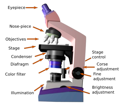
Name and identify the major parts of the microscope. Most are familiar with a light microscope which can sit on a table and allow use to see objects in extremely vivid detail. You cant see any cell with your naked eye because they are very smaller than what human eyes can see normally.
The nucleus cytoplasm cell membrane chloroplasts and cell wall are organelles which can be seen under a light microscope. Light and Electron Microscopes. A Most light microscopes used in a college biology lab can magnify cells up to approximately 400 times and have a resolution of about 200 nanometers.
Details smaller than this are obscured by effects resulting from the wave nature of light. The main onion cell structures are quite easy to observe under medium magnification levels when using a light microscope. Which structure could you observe with a light microscope.
The electron microscope is required to study the structure of viruses. Can change these to focus in more detail and observe smaller structures. Mirror reflecting light through Illuminates the structure of the cells with the denser parts allowing to provide clarity on edges.
This is the reason why you need to use a microscope to observe a cell. A a ribosome B a Golgi apparatus C a nucleus D an endoplasmic reticulum E a peroxisome 2. Using a light microscope one can view cell walls vacuoles cytoplasm chloroplasts nucleus and cell membrane.
Light microscopes use lenses and light to magnify cell parts. In practical terms bacteria and mitochondria which are about 500 nm 05 μm wide are generally the smallest objects whose shape can be clearly discerned in the light microscope. Staining however usually kills the cells.
The size of viruses ranges from 20 to 400 nm which is too small to be seen with an optical microscope. Light microscopes use lenses and light to magnify cell parts. Below the basic structure is shown in the same animal cell on the left viewed with the light microscope and on the right with the transmission electron microscope.
You can not see the very smallest bacteria viruses macromolecules ribosomes proteins and of course atoms. Can you see chromosomes under light microscope. Which of the following structure can you not commonly see with a light microscope and staining techniques a.
You observe a tiny structure under a microscopeWhat question would you ask and then investigate to determine whether the structure was part of a living thing. However they usually can achieve a maximum of 2000x magnification which is not sufficient to see many other tiny organelles. Your instructor will 1 assign you to a microscope 2 provide you with the proper handling instructions of a microscope and 3 guide you through the correct use of the microscope.
The cells look elongated similar in appearance- color size and shape- have thick cell walls and a nucleus that is large and circular in shape. You can not see the very smallest bacteria viruses macromolecules ribosomes proteins and of course atoms. Light and electron microscopes allow us to see inside cells.
You can see most bacteria and some organelles like mitochondria plus the human egg. Demonstrate proper use of the microscope. The smallest bacteria cant be seen with that magnification.
Histological Techniques 6 Visualization Light Microscope Atlas Of Plant And Animal Histology
Light Microscopes An Overview Sciencedirect Topics

Microscope Microscope Parts Labeled Diagram And Functions


Comments
Post a Comment